Researchers at the Institut Pasteur in association with the CNRS have just shown, in an experimental model, that newly formed neurons in the adult brain can be stimulated by light. A novel technique associating optical and genetic tools allows neurobiologists to render neo-neurons photo-excitable. For the first time they have prompted, observed, and specifically recorded the activity of these new nerve cells in the olfactory system. Using this technique the scientists have revealed the nature of signals emitted from new neurons across neuronal circuits in the brain. This work represents an essential step towards better understanding the role of new nerve cells and in developing therapeutic applications, most notably in the realm of neurodegenerative diseases.
Press release
Paris, december 15, 2011
By introducing and inducing expression of photo-sensitive proteins in new neurons , the scientists have been able to control their activity with the use of luminescent flashes. Using this technique, researchers at the Institut Pasteur and the CNRS have been able to observe, stimulate, and specifically record the activity of new nerve cells. They have brought proof that new neurons formed in the olfactory bulb of the adult brain are integrated into preexisting nervous circuits. They have also shown that, against all expectations, the number of contacts between young cells and their target cells greatly increased over several months.
This work constitutes an essential step in characterizing the functions fulfilled by new neurons. It opens new avenues to investigation for understanding the connectivity between “newly formed” neurons and their host circuits. This is a crucial step on the way to foreseeing the use of stem cells within the framework of new therapeutic protocols for repairing brain damage, notably in the realm of neurodegenerative diseases.
- -
* Researchers have succeeded in inducing the expression of a protein sensitive to blue light, from the unicellular algae of the Chlamydomonas genus, in new neurons. Introduced using viral vectors, this protein is expressed on the surface of the neurons. When exposed to blue light the protein opens and thus creates an influx of ions generating an electric field and activating the cell.
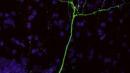
Figure : Neo-neuron marked by the fluorescent protein YFP. © Institut Pasteur
Source
How, when and where new inhibitory neurons release neurotransmitters in the adult olfactory bulb, Journal of Neuroscience, 15 décembre 2010.
Cedric Bardy (1,2), Mariana Alonso (1,2), Walid Bouthour (1,2) et Pierre-Marie Lledo (1,2)
(1) Institut Pasteur, Perception and Memory unit,
(2) CNRS, URA 2182, 25 rue du Dr. Roux, F-75724 Paris Cedex 15, France.
Contact presse
Institut Pasteur Press Office
Sabine D’Andrea - + 33 (0)1 44 38 92 17 - sabine.dandrea@pasteur.fr
Nadine Peyrolo - + 33(0)1 45 68 81 47 - nadine.peyrolo@pasteur.fr