

Researchers at the Institut Pasteur and CNRS, in collaboration with the Synchrotron SOLEIL, have solved the three-dimensional atomic structure of the glycoproteins that envelop the chikungunya virus. This discovery helps to understand how this protein complex is activated in order for the virus to invade target cells. Activation is a key stage in the viral life cycle and understanding its mechanism provides essential information for the development of diagnostic tools, vaccines and potential therapies).
Press release
Paris, december 2, 2010
The atomic structure of the surface protein complex was solved by X-ray crystallography and the placement of the complex into the electron cryomicroscopy map of the virion shows the role it plays in the invasion mechanism. Félix Rey’s team has solved two complexes, each with a specific role in the different stages of the viral life cycle: p62/E1 and E3/E2/E1, the second complex resulting from maturation of the first.
To enter the cell, the virus first uses E2 to bind to the target cell membrane. The membrane then engulfs the virus within vesicles which transport it into the cell through successive compartments. The pH of these compartments (known as endosomes) gradually becomes more acidic, activating E1 which then mediates fusion of the viral and endosomal membranes, enabling the virus to release its RNA into the cell. The RNA is then replicated and used to make viral proteins using the cell machinery leading to the production of infectious viruses which can leave the cell and infect others.
The precursor p62, which is insensitive to acidic pH, binds to E1 and facilitates migration of the complex to the cell membrane. It is during migration that p62 undergoes a maturation process that leads to the creation of proteins E2 and E3. The E3/E2/E1 complexes thus formed assemble to form new viral particles budding from the surface of the infected cell in order to invade new cells.
Understanding these mechanisms is an important step in identifying therapeutic targets. It shows that stabilization of the E3/E2/E1 complex would prevent the virus from invading the cell. The research also identified areas on E2 that recognize neutralizing antibodies, paving the way for new vaccine and diagnostic approaches.
The chikungunya virus, transmitted to humans when bitten by mosquitoes of the genus Aedes, causes severe joint pain in patients. Today medical treatment consists of prescribing anti-inflammatory drugs and painkillers. The epidemic in the Indian Ocean affected almost 2 million people in India and close to 270,000 on the island of Reunion. A new outbreak of the disease occurred in Spring 2010 and the first two indigenous cases were reported in France in September 2010.
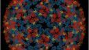
Figure. The surface of a chikungunya virus particle consisting of 240 E3/E2/E1 protein complexes, shown in blue/red/yellow, respectively. These complexes are arranged in a symmetrical pattern around the viral membrane.
This work was funded by the European Community, Actions Marie Curie, INTRAPATH Early Stage Training Program, the Institut Pasteur Transversal Research Program and the Bill & Melinda Gates Foundation.
Source
Glycoprotein organization of chikungunya virus particles revealed by X-ray crystallography, Nature, published on December 2, 2010.
James E. Voss (1,2), Marie-Christine Vaney (1,2), Stéphane Duquerroy (1,2,3), Clemens Vonrhein (4), Christine Girard-Blanc (5,6), Elodie Crublet (5,6), Andrew Thompson (7), Gérard Bricogne (4) and Félix A. Rey (1,2).
(1) Institut Pasteur, Département de Virologie, Unité de Virologie Structurale, 25 rue du Dr Roux, 75724 Paris Cedex 15, France.
(2) CNRS URA 3015, 25 rue du Dr Roux, 75724 Paris Cedex 15, France.
(3) Université Paris-Sud, Faculté d’Orsay, 91405 Orsay Cedex, France.
(4) Global Phasing Ltd, Sheraton House, Castle Park, Cambridge CB3 0AX, United Kingdom.
(5) Institut Pasteur, Département de Biologie Structurale et Chimie, Plateforme de Production de protéines recombinantes, 25 rue du Dr Roux, 75724 Paris Cedex 15, France.
(6) CNRS URA 2185, 25 rue du Dr Roux, 75724 Paris Cedex 15, France.
(7) Synchrotron SOLEIL, L’Orme de Merisiers, BP 48 St Aubin, 91192 Gif sur Yvette, France.
Contact
Institut Pasteur Press Office
Marion Doucet - + 33 (0)1 45 68 89 28 - marion.doucet@pasteur.fr
Nadine Peyrolo - + 33 (0)1 45 68 81 47 - nadine.peyrolo@pasteur.fr

