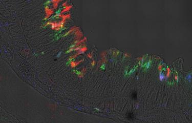
Scientists have assessed the influence of mechanical contractions in the gut on the invasion of two pathogens, one responsible for amebiasis and the other for shigellosis. Their research was based on 3D computational imaging and organ-on-chip technology, which enabled them to accurately reproduce the physiological characteristics of the gut on a chip no larger than a few centimeters.
Physical forces are essential to biological function, but their impact at the tissue level is not fully understood. The gut is under continuous mechanical stress because of peristalsis, a term that refers to all the muscular contractions that enable content to progress through a hollow organ.
At the Institut Pasteur, Nathalie Sauvonnet heads the Intracellular Trafficking and Tissue Homeostasis Group. Elisabeth Labruyère is a scientist in the Biological Image Analysis Unit led by Jean-Christophe Olivo-Marin. The two scientists combined the skills of their respective units to assess the influence of peristalsis on two pathogens in the gut.
- The amoeba Entamoeba histolytica (responsible for amebiasis)
Amebiasis is caused by the amoeba Entamoeba histolytica, a parasite specific to humans. Although infection is generally asymptomatic, if the parasite crosses the gut mucosa it can cause painful, bloody diarrhea and ulcers. In more severe forms it can lead to abscesses in the liver, lungs and brain.
- The bacterium Shigella flexneri (responsible for shigellosis)
Shigellosis is caused by Shigella bacteria, which are specialized clones of Escherichia coli. They contain a virulence plasmid that enables them to invade intestinal epithelial cells and then mucosa. This process results in intense inflammation combined with severe tissue destruction.
Find out more about shigellosis
Reproducing a gut on a chip with gut-on-a-chip technology
Organ-on-chip technology involves accurately reproducing the physiological characteristics of an organ on a cartridge no larger than a USB drive (see inset below: "Organ-on-chip: a technology ripe for further development"). "For our research on peristalsis, organ-on-chip technology enabled us to recreate a sort of functional mini-gut – or gut-on-a-chip," explains Nathalie Sauvonnet. "The two pathogens in this study cause disease only in humans, and no animal model replicates these diseases well." The scientists placed the gut cells under study in the gut-on-a-chip, reproducing the mechanical conditions of a gut using the technology embedded in the chip.
Using gut-on-a-chip and 3D imaging to understand infectivity
The teams led by Nathalie Sauvonnet and Elisabeth Labruyère used gut-on-a-chip technology and computational imaging to study the behavior of the two pathogens. After infecting the device with one of the two microbes, its behavior was imaged in real time in 3D. But confocal microscopes are too slow to image peristaltic motion in the 3D chip. "To observe pathogen invasion in 3D, we had to develop a post-processing method that reconstructs 3D videos by combining 2D acquisitions taken with a programmable microscope. This enabled us to study the infection quantitatively, for example using algorithms to measure the moment at which certain virulence genes are activated," explains Aleix Boquet-Pujadas, lead author of the study. Using the data collected in real time, the authors then examined the role of mechanical stress in infection using a computational method to measure local tension in the tissue under peristalsis. By combining these interdisciplinary approaches involving engineering, biology, physics and quantitative analysis, the scientists obtained a more comprehensive picture of pathogen invasion mechanisms.
This study demonstrates in particular that peristalsis:
- facilitates penetration of the parasite E. histolytica followed by destruction of gut tissue;
- accelerates colonization by S. flexneri and triggers the expression of virulence genes in these bacteria.
These two phenomena are amplified in areas of the tissue that are subject to greater local mechanical stress.
"Our research highlights the fundamental role of physical cues in host-pathogen interactions and introduces a framework that opens the door for mechanobiological research on deformable tissues," explains Elisabeth Labruyère about the study published on October 21, 2022.
Organ-on-chip: a technology ripe for further development
At the Institut Pasteur, the Biomaterials and Microfluidics Platform is rolling out organ-on-chip technology developed by biotech firm Emulate® (Boston, United States). "This technology accurately reproduces the physiological characteristics of different tissues or organs, such as the gut, the pulmonary alveoli and the liver, on a microfluidic chip," explains Samy Gobaa, head of the Institut Pasteur platform. Using organ-on-a-chip makes it possible to act on the three items that aim to avoid animal experimentation or at least to Refine, Reduce and Replace the use of animals in research as soon as possible (the 3Rs guiding principles), which is a commitment for the Institut Pasteur.
Since 2019, this Institut Pasteur organ-on-chip center has launched a series of calls for proposals to facilitate access to the technology.
The Biomaterials and Microfluidics Platform is supported by the Pasteur Microbes and Health Carnot Institute (Pasteur MS).
See also: The potential of "organ-on-chip" technology for biomedical research


